The shell makes the difference

(PhysOrg.com) -- Contrast echography is a commonly used medical imaging technique that is used to show up abnormal blood circulation in organs and tumours. The method makes use of ultrasound and a contrast agent containing special microbubbles. The bubbles are coated with a thin protective layer to prevent them from dissolving in the blood. Until now it was assumed that this layer limited the echo, but now Marlies Overvelde of the University of Twente, The Netherlands, has shown that it is precisely the shell that determines the sensitivity of contrast echography. Her discovery brings the prospect of new diagnostic techniques.
Abnormalities in the blood circulation within organs such as the heart, liver and kidneys can be revealed with the use of contrast echography. In this technique the patient receives an injection of a contrast agent, containing microscopically small bubbles, into the bloodstream. Ultrasound is then transmitted into the body, and the characteristic echo of the bubbles can be picked up efficiently between the surrounding tissues, making it possible to locate the bubbles in the body.
To prevent the bubbles from dissolving in the blood they are coated with an ultra-thin protective layer of lipids. Until now it was always assumed that this layer limited the echo. However, Marlies Overvelde of the University of Twente has discovered that it is precisely this shell that determines the behaviour of contrast bubbles. Moreover, she discovered that the behaviour of medical bubbles used for ultrasound imaging is surprisingly different when they are in the vicinity of a vascular wall.
High speed camera
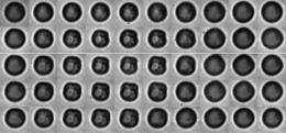
Overvelde made her discovery by using optical laser tweezers to isolate a single contrast bubble, smaller than a red blood cell, from nearby vascular walls and other bubbles. By carefully varying the pressure and frequency of the transmitted ultrasound around the resonance frequency, she could record the bubble oscillations with a high speed camera capable of recording 25 million images per second. 'In this way you can see how the bubble reacts to ultrasound with amazing accuracy,' says Overvelde. 'The nanometer-thin shell buckles as the bubble shrinks and ruptures as the bubble grows.' Overvelde observed the very same non-linear properties of the bubble shell in the characteristic response of the bubble. This explains the sensitivity of contrast echography.
The position of the bubble could also be manipulated with the optical tweezers. The behaviour of the bubble was shown to change when it was close to a wall or to other bubbles. 'These observations are broadly in line with what we already know about larger bubbles, but we are discovering exciting new details about the interaction between bubbles at the microscopic level; a field that is largely unexplored territory within physics.'
New diagnostic techniques
The discovery opens the way for new diagnostic techniques, including imaging at the molecular level. For this, microbubbles will be coated with biochemical ligands that attach them to diseased or pathogenic cells. The researcher hopes that the method can be used to detect illnesses in their very early stages using relatively simple and inexpensive ultrasound imaging.
The research was carried out as part of a major strategic project of the European Commission, in which a consortium from seven European countries (made up of companies in the medical technology and pharmaceutical industries, academic research institutions and medical centres) is working on methods for detecting prostate cancer at an early stage. The project is closely related to the groundbreaking NIMTIK research of the University of Twente, which is working on increased contrast for the optical and acoustic techniques used to localize and kill tumours at an early stage.
Overvelde received her PhD from the faculty of Science and Technology on 9 April.
Provided by University of Twente


