CT with Nanotubes for People and Baggage Scans
Researchers from Siemens are investigating the use of small, fast X-ray sources based on nanotubes.
In combination with a computer tomograph (CT) scanner, these could serve to generate high-quality images of rapid processes within the human body, such as the dispersion of a contrast medium. The radiation would be reduced without any sacrifice of image quality compared to the equipment in use today. The use of fast X-ray sources is also attractive for applications requiring a fast throughput. These include scanning systems for luggage and passengers in airports. Siemens is developing these sources together with the U.S. company Xintek in a joint venture by the name of XinRay Systems.
X-rays are generated when accelerated electrons strike an electrode. Today's conventional source for such electrons is a hot filament inside a vacuum tube. However, this type of system consumes a lot of energy, is reacts relatively slow, gets hot, and can only be miniaturized to a certain degree. Therefore, researchers look for "cold" electron sources which emit electrons under high voltage from a metal formed into tiny spikes or sharp edges.
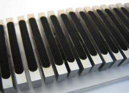
Nanotubes made of carbon are ideal as so-called field emitters, since they are as conductive as metal and also very thin, with a diameter of a few nanometers. They can be activated in less than a millionth of a second, whereas it takes several hundredths of a second to trigger a conventional X-ray source. The researchers at Siemens, Xintek and XinRay first apply nanotubes to a metal substrate and then control these individually in order to create an array of mini electron sources. The technology stems from research by a team at the University of North Carolina and is now being developed into a commercial solution by Siemens.
In the most powerful CT scanners currently available from Siemens, two X-ray tubes revolve around the patient at more than three times per second. In the future, hundreds of mini X-ray sources could be permanently installed in a fixed circle and triggered in sequence. This would generate ten high-quality images per second — fast enough to enable observation of processes such as the destruction of tumor tissue during radiation therapy. It will take several years before such X-ray sources can be used in series-produced medical devices. However, their market readiness for use in industrial environments or to scan baggage in airports could come much sooner.
Source: Siemens


