'Nanodrop' Test Tubes Created with a Flip of a Switch

Researchers at the National Institute of Standards and Technology have demonstrated a new device that creates nanodroplet “test tubes” for studying individual proteins under conditions that mimic the crowded confines of a living cell. “By confining individual proteins in nanodroplets of water, researchers can directly observe the dynamics and structural changes of these biomolecules,” says physicist Lori Goldner, a coauthor of the paper published in Langmuir.
Researchers recently have turned their attention to the role that crowding plays in the behavior of proteins and other biomolecules—there is not much extra space in a cell. NIST’s nanodroplets can mimic the crowded environment in cells where the proteins live while providing advantages over other techniques to confine or immobilize proteins for study that may interfere with or damage the protein.
This more realistic setting can help researchers study the molecular basis of disease and supply information for developing new pharmaceuticals. For example, misfolded proteins play a role in many illnesses including Type 2 diabetes, Alzheimer’s and Parkinson’s diseases. By seeing how proteins fold in these nanodroplets, researchers may gain new insight into these ailments and may find new therapies.
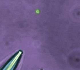
The NIST nanodroplet delivery system uses tiny glass micropipettes to create tiny water droplets suspended in an oily fluid for study under a microscope. An applied pressure forces the water solution containing protein test subjects to the tip of the micropipette as it sits immersed in a small drop of oil on the microscope stage. Then, like a magician whipping a tablecloth off a table while leaving the dinnerware behind, an electronic switch causes the pipette to jerk back, leaving behind a small droplet typically less than a micrometer in diameter.
The droplet is held in place with a laser “optical tweezer,” and another laser is used to excite fluorescence from the molecule or molecules in the droplet. In one set of fluorescence experiments, explains Goldner, “The molecules seem unperturbed by their confinement—they do not stick to the walls or leave the container—important facts to know for doing nanochemistry or single-molecule biophysics.” Similar to a previous work (see “‘Micro-boxes’ of Water Used to Study Single Molecules”, Tech Beat July 20, 2006), researchers also demonstrated that single fluorescent protein molecules could be detected inside the droplets.
Fluorescence can reveal the number of molecules within the nanodroplet and can show the motion or structural changes of the confined molecule or molecules, allowing researchers to study how two or more proteins interact. By using only a few molecules and tiny amounts of reagents, the technique also minimizes the need for expensive or toxic chemicals.
Citation: J. Tang, A.M. Jofre, G.M. Lowman, R.B. Kishore, J.E. Reiner, K. Helmerson, L.S. Goldner and M.E. Greene. Green fluorescent protein in inertially injected aqueous nanodroplets. published in Langmuir, ASAP Article, Web release date: March 27, 2008.
Source: National Institute of Standards and Technology


