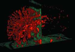
New imaging technique allows doctors to 'see' molecular activity

A new technique that will enable doctors to ‘see’ things happening at the molecular level using standard imaging techniques has been developed by Oxford scientists. The technique has initially been directed towards multiple sclerosis, but long-term it has the potential to be used for a vast range of health problems. The findings are published by Nature Medicine on Monday 24 September.
Dr Robin Choudhury and colleagues at the University of Oxford have developed a marker that attaches itself to particular molecules involved in inflammation. As a result, these molecules ‘light up’ on MRI scans.
The ‘VCAM-1’ molecule plays a key role in inflammation, which contributes to many diseases, including multiple sclerosis, arthritis, inflammatory bowel disease and atherosclerosis (a hardening and narrowing of the arteries which can lead to heart attack and stroke).
By injecting into the body markers that attach themselves to VCAM-1 molecules, and which are visible under MRI scanning, the researchers were able to see on a scan exactly where the molecules were in operation, and in what quantities. The ultimate goal is to facilitate earlier diagnosis, guide treatment and provide more precise monitoring of disease progression.
The team has developed the technique with a view to using it in multiple sclerosis (MS), an autoimmune disease that affects the central nervous system. In MS the body’s own immune system, through inflammation, attacks the fatty tissue surrounding nerve fibres (myelin), which helps nerves carry signals, leading to a range of problems with vision and movement.
Compared to conventional MRI techniques, the new technique has the potential to reveal disease activity much earlier and, crucially, before tissue destruction has occurred. Earlier intervention with drug treatments guided by this information may alter disease progression.
‘Ordinarily, by the time lesions are visible, damage is already done,’ says Dr Choudhury. ‘Therefore molecular imaging techniques to accelerate diagnosis are urgently needed, and we think that this approach shows great potential.’
The work has been carried out so far in mice, but the researchers are optimistic that the technique could translate to humans.
‘In the last few years our knowledge about the cellular and molecular basis of diseases, including MS, has expanded vastly, but we haven’t yet matched these with accompanying advances in imaging,’ says Dr Choudhury. ‘The new technique helps to address that issue.
‘The ultimate goal of this line of work is to develop tools than can accelerate diagnosis, guide specific therapies and monitor response to treatment. In the paper, we make a case as to how this could be applied to multiple sclerosis, but in fact this is a platform technology that, in theory, could be adapted for diagnosis in other brain diseases, cancer and coronary artery disease.’
The full paper ’In vivo magnetic resonance imaging of acute brain inflammation using microparticles of iron oxide’ appears online at www.nature.com/nm/
Source: University of Oxford


