Unprecedented 3-D images of human ear anatomy for hearing restoration

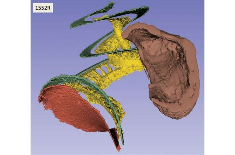
Using advanced techniques at the Canadian Light Source (CLS) at the University of Saskatchewan, scientists have created three-dimensional images of the complex interior anatomy of the human ear, information that is key to improving the design and placement of cochlear implants.
"With the images, we can now see the relationship between the cochlear implant electrode and the soft tissue, and we can design electrodes to better fit the cochlea," said Dr. Helge Rask-Andersen, senior professor at Uppsala University in Sweden.
"The technique is fantastic and we can now assess the human inner ear in a very detailed way."
The cochlea is the part of the inner ear that looks like a snail shell and receives sound in the form of vibrations. In cases of hearing loss, cochlear implants are used to bypass damaged parts of the ear and directly stimulate the auditory nerve. The implant generates signals that travel via the auditory nerve to the brain and are recognized as sound.
By imaging the soft and bony structures of the inner ear with implant electrodes in place, Rask-Andersen said the researchers were able to discover what the auditory nerve looks like in three dimensions, and to learn how cochlear implant electrodes behave inside the cochlea. This is very important when cochlear implants are considered for people with limited hearing.
"When we operate on patients with some residual hearing, we must be extremely atraumatic; the electrode must be very soft and inserted with no trauma. Its location inside the cochlea must be optimal, meaning it should not perforate any membranes and it should also be away from the vibrating membrane filtering the low frequencies that are often preserved in the patient."
The research, conducted with colleagues from Western University and published in Ear and Hearing, the official journal of the American Auditory Society, provides information that can be used to assess electrode insertion depths and stimulation strategies as well as to create exact frequency maps for optimal stimulation of the auditory nerve.
Detailed visualization of the inner ear also creates opportunities to explore potential remedies for other afflictions, he said.
One that is of particular interest to Rask-Andersen is Meniere's disease, which causes pressure, severe dizziness, hearing loss and a ringing or roaring noise.
"The next steps in the research will be to look at the anatomy of the fluid-draining pathways important to understanding Meniere's disease," said Rask-Andersen.
More information: Hao Li et al. Synchrotron Radiation-Based Reconstruction of the Human Spiral Ganglion, Ear and Hearing (2019). DOI: 10.1097/AUD.0000000000000738
Provided by Canadian Light Source


