Visualization of the behavior of sugar transport proteins

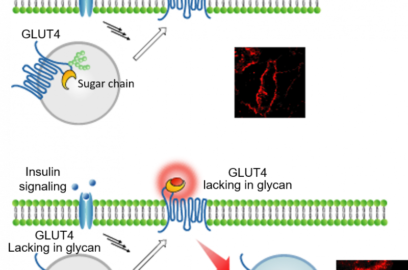
A group of researchers led by Kazuya Kikuchi, professor at the Graduate School of Engineering, Osaka University, clarified the role of a N-glycan chain on glucose transporter type 4 (GLUT4) by developing a method for visualizing intracellular trafficking of proteins.
The GLUT4 translocation disorder is known to be associated with the onset of type II diabetes. GLUT4 is translocated to the cell membrane in response to insulin stimulation and takes up glucose in blood, decreasing blood sugar levels. The role of the N-glycan chain on GLUT4 in intracellular trafficking has drawn attention in recent years.
The fluorogenic probe developed by this group has the function of increasing fluorescence intensity by binding to a protein. Thanks to the cell-impermeability of the fluorogenic probe, it's possible to label only proteins that appear on the cell membrane. These functions have made it possible to quickly fluorescence-label only GLUT4 that is translocated to the cell membrane by fluorogenic probes and clearly detect fluorescence of GLUT4 translocation to the cell membrane.
Previously, kinetic analysis of GLUT4 was performed by using fluorescence proteins and an immunostaining method; however, it was impossible to precisely examine if GLUT4 in the cell had been transiently translocated to the cell membrane.
The technique developed by this group has made it possible to precisely determine the translocation progress of GLUT4; that is, it has become possible to record the evidence showing that GLUT4 was transiently translocated to the cell membrane. It was clarified that GLUT4 with abnormalities in the N-glycan chain was transiently translocated to the cell membrane but was rapidly internalized without retention on the cell membrane, and that the N-glycan chain played a role in retaining GLUT4 on the cell membrane.
This group's achievement will lead to the clarification of the mechanism behind the localization of GLUT4 and the mechanism for developing diabetes, as well as the development of new types of treatment drugs.
More information: Shinya Hirayama et al. Fluorogenic probes reveal a role of GLUT4 N-glycosylation in intracellular trafficking, Nature Chemical Biology (2016). DOI: 10.1038/nchembio.2156
Journal information: Nature Chemical Biology
Provided by Osaka University


