Seeing DROSHA for the first time: Lab team gets the first glimpse of elusive protein structure

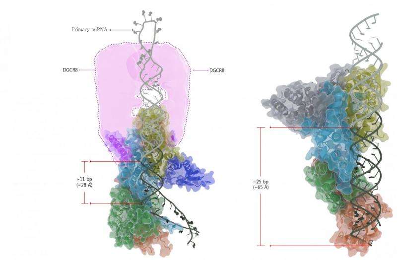
Our bodies are made up of many different types of cells, with each of their identities determined by different gene expression. Cancer and genetic diseases occur when this gene expression goes wrong. MicroRNAs (miRNAs) are an important regulator in gene expression, and they play crucial roles in almost all biological contexts including development, differentiation, inflammation, aging, and cancer. In the nucleus, miRNA start their process as a tiny, folded over hairpin structure called primary microRNA (pri-miRNA) and is recognized and processed by the Microprocessor complex, an enzyme arrangement made up of one DROSHA and two DGCR8 proteins. The Microprocessor complex does two things: it measures the pri-miRNA then snips off its basal parts, resulting in precursor-microRNA (pre-miRNA). After some further processing, mature miRNA and RNA-Induced Silencing Complex (RISC) interact with messenger RNA (mRNA) in the cytoplasm to repress translation which stops the ribosome from making protein.
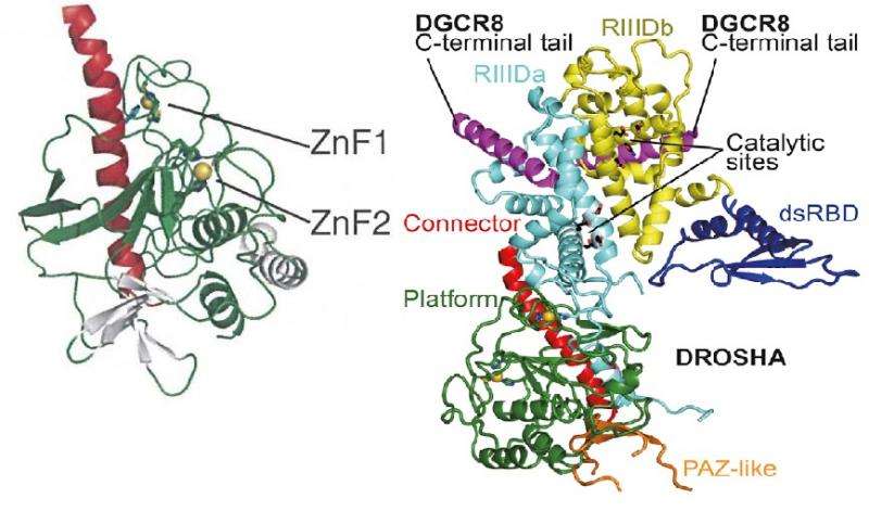
While this process has been studied thoroughly over the last decade, the molecular basis of Microprocessor is still poorly understood because the structures of the proteins have not been sufficiently revealed yet. Scientists working at the IBS Center for RNA Research in South Korea are at the forefront of discovering and mapping out the complexities of the structures involved in this process of pri-miRNA biogenesis. A team led by V. Narry Kim has for the first time elucidated a three dimensional image of DROSHA, one part of the Microprocessor complex. Until now, no one has been able to obtain DROSHA's crystal structure.
With this discovery, the IBS team was able to confirm their previous findings (Cell Nguyen et al., 2015) which revealed the composition and detailed action mechanism of the Microprocessor complex. This work showed that DROSHA has two DGCR8-binding sites, giving a clear picture of how Microprocessor is assembled. After understanding its structure they were able to determine how DROSHA, along with DGCR8, interact together to determine the cleavage sites in pri-miRNA. Previously they observed that Microprocessor complex cuts the middle stem region of the pri-miRNA at a distance of 11 base pairs from the basal junction. The current research helped them to determine that the shape of DROSHA has unique physical characteristics, including a "bump", which may accommodate pri-miRNA perfectly. This bump may act as a measuring guide and indicates the 11-base-pair distance for DROSHA to cleave.

When they looked at DROSHA, they noticed that it bore some striking structural similarities to the Dicer enzyme, despite Dicer existing and functioning away from DROSHA. They have hypothesized that DROSHA may have evolved from a Dicer homologue.
Attempts to purify the over-expressed DROSHA protein for study have previously been impossible and have been hindered by technical difficulties. Without exacting care, DROSHA is easily aggregated during a protein purification process, thereby becoming unsuccessful crystallization. To maintain its structural integrity, the team co-expressed 23 amino acids from DGCR8 which binds and covers hydrophobic surface of DROSHA, keeping DROSHA intact. According to IBS researcher Jae-Sung Woo, "Without this hydrophobic interaction, the DROSHA proteins will fold abnormally and aggregate," thus rendering them useless. Because the proteins were maintained, the team was able to perform x-ray crystallography and get the first clear image of their structure.
Understanding DROSHA's structure is another crucial step in the process of understanding microRNA biogenesis. Building on this knowledge is the basis for devising new ways of regulating and controlling gene expression, which has a range of applications including building new antifungals and stopping tumor growth. Being able to more precisely control RNAi may be on the forefront of combating otherwise untreatable bacterial infections as antibiotics resistance grows. Achieving this imaging breakthrough opens the door for a better understanding and exciting new applications in cell reproduction. "In the future," says researcher Sung Chul Kwon, "we are planning to solve the structure of pri-miRNA-bound Microprocessor complex."
More information: S. Chul Kwon, Tuan Anh Nguyen, Yeon-Gil Choi, Myung Hyun Jo, Sungchul Hohng, V. Narry Kim, Jae-Sung Woo; Structure of Human DROSHA; Cell, DOI: dx.doi.org/10.1016/j.cell.2015.12.019
Journal information: Cell
Provided by Institute for Basic Science


