Metabolite maps provide more information and can help reduce harvest losses
It is possible to make metabolite maps of living plant tissues both quickly and accurately by using laser ablation electrospray ionization mass spectrometry imaging. Researchers at Wageningen UR describe this in their article in the scientific journal Plant Physiology. The maps are 2-D at present, but the researchers are planning to investigate whether they can also use the technique to make 3-D maps.
The maps the Wageningen researchers have made are accurate to half a millimetre and can be used, for example, to improve our understanding of how plants react to invading pathogens. Such information is important for developing food crops which will be more resistant to diseases and plagues and produce better food as a result.
More than 100,000 plant compounds
Every plant cell contains thousands of metabolites. Cell enzymes make the production of all these metabolites possible. The presence of the enzymes is in turn controlled by genes in the DNA of the plant. The active genes in petals are different to those, for example, in green leaves. This means that metabolites responsible for giving a flower its colour are only made in petals. All the thousands of metabolites in plant cells have different functions: the pigments in a flower ensure pollinating insects are enticed to visit it; particular flavours make berries attractive to birds, so that the latter can spread the seeds; and alkaloids and many other specialized metabolites help plants defend themselves against diseases and pests.
There are often huge differences between the composition of plant cells situated close to each other, for example, inside a leaf. That is why it is useful to be able to measure the compounds in specific groups of cells, or even individual cells. There was already a technique for doing this in dried tissues but it is less reliable because the composition of cells can change during the drying process. In other words, it is much better to measure living tissues.
Lasers, explosies en elektrische lading
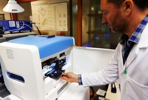
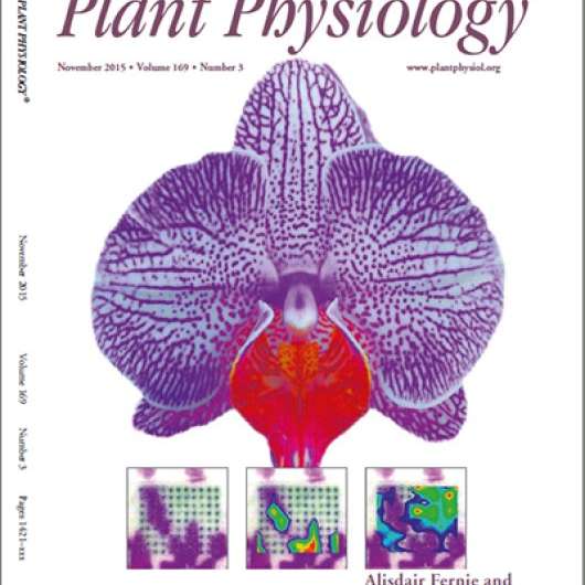
The Wageningen researchers looked at whether laser ablation electrospray ionization mass spectrometry imaging equipment, provided by CAT–AgroFood shared research facilities at Wageningen UR, could be used to make detailed metabolite maps. They used orchid flowers to test this. The Phalaenopsis orchid flowers have varieties which are white, with purple speckles. First of all, purple and white tissues were investigated in a traditional way by separating out the two colour tissues using a scalpel and forceps. Then a standard analysis was also used to determine which substances were present in each of the two tissues. In this way it was possible to identify the substances from the white and the purple tissues.
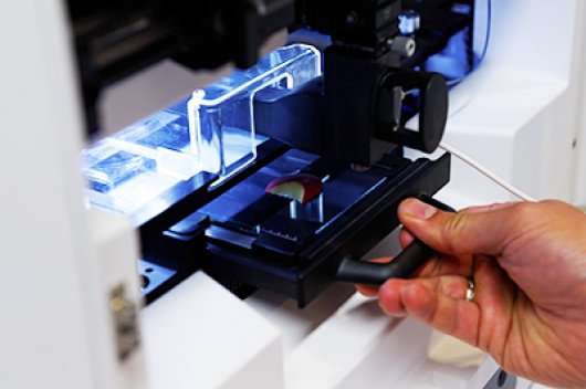
Subsequently, laser ablation electrospray ionization was used on petals. In this technique the laser is directed onto a small group of cells. The intensity of the laser beam is adjusted so that it makes the water in the cells boil within a few milliseconds, causing the cells to explode. This releases all the metabolites into the air as tiny droplets just above the petal, forming a solvent mist. The released metabolites become electrically charged at the same time, and are drawn into the mass spectrometer because of the presence of an electric field. In this mass spectrometer the size of the molecules can be measured very accurately. This makes it possible to determine which metabolites occur at which specific points in the plant tissue.
Mapped down to the millimetre
If the laser is coupled to a computer, a 'grid' of measuring points can be investigated. The Wageningen researchers, having chosen a distance of half a millimetre between measuring points, could analyze hundreds of groups of cells in the petals in just a few minutes.
By comparing results with earlier analyses of purple and white petal tissues, they could prove that this technique really does work. The compounds found with the new technique only when the laser hit a group of purple cells were also found only in the purple tissues using the standard analysis. Similarly, the metabolites found using the new technique when the laser hit a group of white cells were also the ones the traditional analysis identified in white tissues.
The maps are now in 2-D format, but the researchers are investigating whether they can 'look' more deeply into plant tissues, so that even 3-D maps can be made.
Knowledge leading to more and better food
The functions of more and more metabolites in plants are being discovered. But where and how they function has been difficult to investigate up until now. For example, we know that certain metabolites ensure plants can defend themselves against diseases and pests. Until now, such substances could only be investigated by using tissue pieces from diseased and healthy leaves, and then comparing the results. The Wageningen researchers have now shown that it is also possible to make an accurate map of the defence reaction directly in living leaves. This new technique can teach us far more about the often very localized interactions between plants and their attackers. This knowledge can be used to develop crops that can defend themselves better against diseases and plagues and therefore produce more and better foods.
More information: Spatially Resolved Plant Metabolomics: Some Potentials and Limitations of Laser-Ablation Electrospray Ionization Mass Spectrometry Metabolite Imaging. Plant Physiology November 2015 doi: dx.doi.org/10.1104/pp.15.01176
Provided by Wageningen University


