New approach to mammograms could improve reliability

Detecting breast cancer in women with dense mammary tissues could become more reliable with a new mammogram procedure that researchers have now tested in pre-clinical studies of mice. In their report in the journal ACS Nano, they describe injecting gold nanoparticles in mammary tissue to enhance the imaging of early signs of breast cancer.
Mammography remains the clinical gold standard of screening tests for detecting breast cancer. However, a recognized limitation of this X-ray procedure is that dense breast tissue shows up as white masses and fibers on an image, which can obscure the presence of microcalcifications—potential signs of early cancer development. Other imaging methods including ultrasound, magnetic resonance imaging and molecular breast imaging can also find abnormalities, but each has its own limitation, such as high cost or poor resolution. Lisa Cole, Tracy Vargo-Gogola and Ryan K. Roeder wanted to improve patients' options.
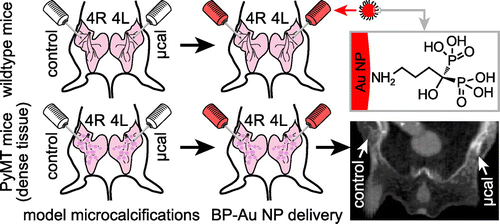
The researchers boosted the contrast of mammography X-rays by modifying gold nanoparticles with molecules that bind specifically to microcalcifications. They injected a low dose of these nanoparticles into the mammary glands of mice with dense tissue. The engineered particles made the microcalcifications brighter on the X-rays—and therefore, easier to distinguish. The mice showed no obvious side effects. Although further research would be required, the scientists say the technique could eventually translate into more reliable breast cancer detection for women with dense mammary tissue.
More information: Contrast-Enhanced X-Ray Detection of Microcalcifications in Radiographically Dense Mammary Tissue Using Targeted Gold Nanoparticles, ACS Nano, Article ASAP, DOI: 10.1021/acsnano.5b02749
Abstract
Breast density reduces the accuracy of mammography, motivating methods to improve sensitivity and specificity for detecting abnormalities within dense breast tissue, but preclinical animal models are lacking. Therefore, the objectives of this study were to investigate a murine model of radiographically dense mammary tissue and contrast-enhanced X-ray detection of microcalcifications in dense mammary tissue by targeted delivery of bisphosphonate-functionalized gold nanoparticles (BP-Au NPs). Mammary glands (MGs) in the mouse mammary tumor virus - polyomavirus middle T antigen (MMTV-PyMT or PyMT) model exhibited greater radiographic density with age and compared with strain- and age-matched wild-type (WT) controls at 6–10 weeks of age. The greater radiographic density of MGs in PyMT mice obscured radiographic detection of microcalcifications that were otherwise detectable in MGs of WT mice. However, BP-Au NPs provided enhanced contrast for the detection of microcalcifications in both radiographically dense (PyMT) and WT mammary tissues as measured by computed tomography after intramammary delivery. BP-Au NPs targeted microcalcifications to enhance X-ray contrast with surrounding mammary tissue, which resulted in improved sensitivity and specificity for detection in radiographically dense mammary tissues.
Journal information: ACS Nano
Provided by American Chemical Society


