Novel probe for live human iPS cell imaging

Researchers from the National Institute of Advanced Industrial Science and Technology (AIST) have developed a highly sensitive lectin probe, rBC2LCN, for human induced pluripotent stem cells (iPS cells). rBC2LCN allows staining of live iPS cells just by adding the probe to an iPS cell culture medium. The researchers have also revealed that rBC2LCN binds to H-type 3 O-glycans on membrane proteins of iPS cells.
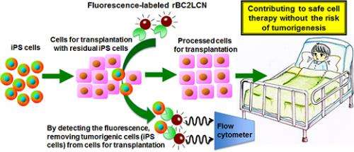
rBC2LCN can evaluate the undifferentiated status of iPS cells, allowing quality control and more efficient culture of iPS cells. A major subject for the use of iPS cells in regenerative medicine is that residual iPS cells in transplanting differentiated cells could form tumors. The rBC2LCN probe would contribute to safe cell transplantation by allowing visualization and removal of the residual iPS cells.
The technology will be presented in detail on March 22, 2013, at the 12th Congress of the Japanese Society for Regenerative Medicine to be held in Yokohama, Kanagawa Prefecture. The technology will be published online in a U.S. journal, Stem Cells Translational Medicine.
In recent years, development of a system to ensure a stable supply of quality controlled iPS cells in large quantities has become important in response to the high expectations held for regenerative medicine using human stem cells such as iPS cells. To keep quality of iPS cells, they need to be efficiently distinguished from other cells. There are several cell staining methods including methods for formalin-fixed and live iPS cells, but they are inadequate in terms of sensitivity and other aspects. Therefore, there is a strong need to develop an undifferentiation marker that can determine the quality of iPS cells in easy and sensitive manners.

A number of researchers have been working on regulation of differentiation into various cells from iPS cells, such as neurons and retinal cells, to realize cell therapy. A major subject for preparing differentiated cells from iPS cells is residual undifferentiated iPS cells, which have tumorigenic potential. This is a major obstacle to the application of iPS cells to regenerative medicine. Therefore, a novel probe is required, which could remove residual iPS cells completely.
AIST aims to be a front runner in the industrial application of stem cells, with a particular focus on stem cell differentiation technology and stem cell standardization. AIST has also been working on the application of a high-density lectin microarray as a novel technology for glycan analysis. AIST has developed an excellent staining probe of iPS cells without the use of conventional antibodies by combination of these technologies.

This research was conducted by AIST as a project (FY2009 - 2010) supported by the New Energy and Industrial Technology Development Organization and a collaborative research of AIST and Wako Pure Chemical Industries (from FY2011) funded by Wako Pure Chemical Industries.
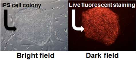
A high-density lectin microarray analysis revealed that rBC2LCN, a lectin probe, binds specifically to iPS cells. The researchers found that rBC2LCN is effective for staining of live iPS and embryonic stem (ES) cells. Figure 1 shows the results of staining of live iPS cells with rBC2LCN. Live iPS cells could be fluorescently stained by adding rBC2LCN labeled with a red fluorescent dye to the culture medium of the iPS cells. This probe shows no toxicity and can be left in the culture medium, allowing the constant control of iPS cell quality during culture. rBC2LCN is also capable of live cell staining of ES cells, being applicable to quality control of varied stem cells.

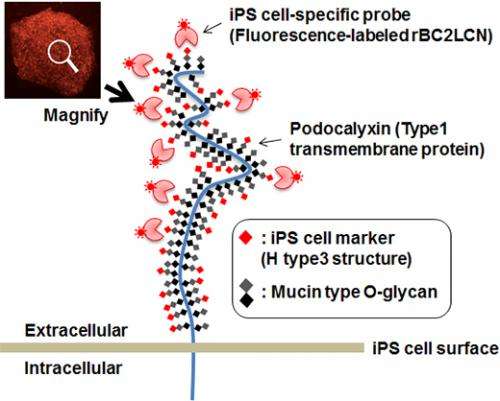
The researchers examined the molecules on iPS cells recognized by rBC2LCN. They demonstrated that rBC2LCN binds to O-glycans on a heavily glycosylated membrane protein called podocalyxin (Fig. 2). rBC2LCN recognizes an H-type 3 structure of O-glycans (Fucα1-2Galβ1-3GalNAc). This suggests that H-type 3 is a novel iPS cell marker.
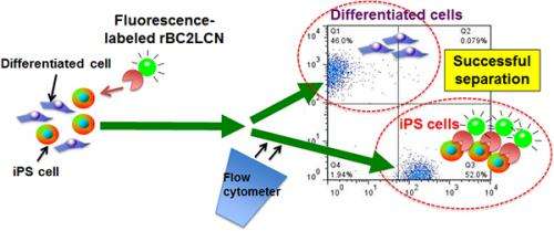
Flow cytometry was conducted after the addition of fluorescence-labeled rBC2LCN to a mixture of cells containing iPS cells and differentiated cells. iPS cells could be separated from the differentiated cells (Fig. 3). Thus, even if iPS cells are present among cells prepared for transplantation in regenerative medicine, it will be possible to prevent tumor formation by using this technology to separate out the iPS cells.
rBC2LCN can be produced in large quantities (>80 mg/L) by using Escherichia coli. Therefore rBC2LCN could be a novel type of iPS cell probe that is cost effective unlike conventional antibodies.
Journal information: Stem Cells Translational Medicine
Provided by Advanced Industrial Science and Technology


