Team develops new method for filming the release of medicine inside cells

Apr 27, 2012

Researchers at the University of Twente (The Netherlands) have developed a method to allow them to watch close-up how medicines are released in cells and to monitor their absorption at the level of individual cells. The method makes use of the fastest camera in the world, which can film at a rate of 25 million images per second. This new method is an important step forward for research into how medicines could be directed to a specific part of the body using microscopic bubbles.

There are various ways in which medicine can be taken – orally, in an injection or through a drip, for example. In all these methods, it is the blood that takes the medicine to its ultimate destination – such as the site of an infection or tumour. But of course, the blood takes the medicine all around the body indiscriminately, which can actually lead to damage elsewhere. The drugs used in chemotherapy, for example, attack both healthy and tumour cells.

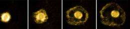
So the University of Twente has been working on new methods for targeting medicines on very specific sites in the body. One of these methods involves using containing medicine. When these bubbles are injected into the patient’s bloodstream, they can be activated using ultrasound when they reach a specific part of the body (such as the site of a tumour). The vibrations cause the bubbles to release the medicine, which can also be absorbed more easily by the cells because the bubbles can actually pass through tiny holes in the cell walls.

This method of administering medicine has considerable potential, but more research is still required. One limiting factor has been that – until now – it was not possible to see how medicines were being absorbed, simply because the whole process happened too quickly and the bubbles were too small to see using normal microscopes. This is why researchers of the University of Twente’s Physics of Fluids research group, together with researchers from the Erasmus MC, set out to develop a new method for viewing the of medicines at the cellular level. The , which has been dubbed ‘ultra-high-speed fluorescence microscopy’, makes use of the Brandaris 128 camera – which was also developed at Twente. The camera can film at a speed of at least 25 million per second, making it the fastest camera in the world. Various other clever adaptations have made this not only the fastest camera in the world, but also one of the most sensitive of its kind.

Explore further: Scientists develop advanced biological computer

More information: Erik Gelderblom of the Faculty of Science and Technology defended his PhD thesis on this subject on Friday 20 April.

add to favorites email to friend print save as pdf

Related Stories

The shell makes the difference

Apr 12, 2010

(PhysOrg.com) -- Contrast echography is a commonly used medical imaging technique that is used to show up abnormal blood circulation in organs and tumours. The method makes use of ultrasound and a contrast ...

Spontaneous combustion in nanobubbles

Sep 28, 2011

(PhysOrg.com) -- Nanometer-sized bubbles containing the gases hydrogen and oxygen can apparently combust spontaneously, although nothing happens in larger bubbles. For the first time, researchers at the University ...

Bursting bubbles with sound offers new treatments for cancer

Jun 28, 2010

A new way to deliver cancer drugs using gas bubbles and sound waves is to be developed at the University of Leeds. The project will enable highly toxic drugs to be delivered in small doses directly to tumours, where their ...

Photoacoustics useful in cancer research

Dec 05, 2008

(PhysOrg.com) -- Photoacoustics can be used to show the development of blood capillaries in and around a tumour. PhD student, Kiran Kumar Thumma, of the University of Twente (Netherlands) is the first to use ...

Opening the brain to new treatments

Mar 13, 2012

One of the trickiest parts of treating brain conditions is the blood brain barrier, a blockade of cells that prevent both harmful toxins and helpful pharmaceuticals from getting to the body's control center. ...

Recommended for you

Scientists develop advanced biological computer

May 24, 2013

(Phys.org) —Using only biomolecules (such as DNA and enzymes), scientists at the Technion-Israel Institute of Technology have developed and constructed an advanced biological transducer, a computing machine capable of manipulating ...

Research aims to fix long-held, inaccurate insect model

May 24, 2013

(Phys.org) —In humans, a polymer called melanin determines skin, eye and hair color—the darker the skin, the more melanin in a person's body. For insects, melanin is a major aspect of their immune defense ...

Molecular modelling to help create better, safer drugs

May 24, 2013

(Phys.org) —How our bodies break down the common drugs ibuprofen, diclofenac and warfarin is the subject of a new study from the University of Bristol, published in the Journal of the American Chemical So ...

User comments : 0

More news stories

New method for producing clean hydrogen

Duke University engineers have developed a novel method for producing clean hydrogen, which could prove essential to weaning society off of fossil fuels and their environmental implications.

Scientists develop advanced biological computer

(Phys.org) —Using only biomolecules (such as DNA and enzymes), scientists at the Technion-Israel Institute of Technology have developed and constructed an advanced biological transducer, a computing machine capable of manipulating ...