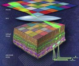
Hi-res detector used by researchers to map neural circuits of the retina

(PhysOrg.com) -- Using a sophisticated neural recording system developed by physicists at UC Santa Cruz, researchers were able to trace for the first time the neural circuitry that connects individual photoreceptors with retinal ganglion cells, the neurons that carry visual signals from the eye to the brain.
Their study, published in the October 7 issue of Nature, describes neural circuits in the retina at the resolution of individual neurons and provides insight into the neural code used by the retina to relay color information to the brain.
"Our neural recording system is able to record simultaneously the tiny electrical signals generated by hundreds of the retinal output neurons that transmit information about the outside visual world to the brain," said coauthor Alan Litke, adjunct professor of physics at UCSC.
Litke and coauthor Alexander Sher, assistant professor of physics, lead the work at UCSC on the project, which involves a large team of collaborators, including biologists at the Salk Institute for Biological Studies.
"Nobody has ever seen the entire input-output transformation performed by complete circuits in the retina at single-cell resolution," said senior author E. J. Chichilnisky of the Salk Institute. "We think these data will allow us to more deeply understand neuronal computations in the visual system and ultimately may help us construct better retinal implants."
The retinal readout device that made the experiments possible was inspired by detector technologies that physicists are using to search for the Higgs boson at the Large Hadron Collider. It was developed by an international team of high-energy physicists from the Santa Cruz Institute for Particle Physics (SCIPP) at UCSC, the AGH University of Science and Technology in Krakow, Poland, and the University of Glasgow, U.K. The system records neural signals at high speed (over ten million samples each second) and with fine spatial detail, sufficient to detect even a locally complete population of the tiny and densely spaced output cells known as "midget" retinal ganglion cells.
Retinal ganglion cells are classified based on their size, the connections they form, and their responses to visual stimulation, which can vary widely. Despite their differences, they all have one thing in common--a long axon that extends into the brain and forms part of the optic nerve.
Visual processing begins when photons entering the eye strike one or more of the 125 million light-sensitive nerve cells in the retina. This first layer of cells, which are known as rods and cones, converts the information into electrical signals and sends them to an intermediate layer, which in turn relays signals to the 20 or so distinct types of retinal ganglion cells.
In an earlier study, the researchers found that each type of retinal ganglion cell forms a seamless lattice covering visual space that transmits a complete visual image to the brain. In the current study--led by Sher and Salk postdoctoral researchers Greg Field and Jeffrey Gauthier--the team zoomed in on the pattern of connectivity between these layers of retinal ganglion cells and the full lattice of cone receptors.
To resolve the fine structure of receptive fields--the small, irregularly shaped windows through which neurons in the retina view the world--the authors used stimuli with tenfold smaller pixels. "Instead of a diffuse region of light sensitivity, we detected punctate islands of light sensitivity separated by regions of no light sensitivity," Field said.
When combined with information on spectral sensitivities of individual cones, maps of these punctate islands not only allowed the researchers to recreate the full cone mosaic found in the retina, but also to conclude which cone fed information to which retinal ganglion cell.
"Just by stimulating input cells and taking a high density recording from output cells, we can identify all individual input and output cells and find out who is connected to whom," Chichilnisky said.
At UCSC, researchers have created several versions of the neural recording system, including advanced versions that are able to both stimulate and record neural activity in a large population of neurons. Sher, Litke, and their collaborators are using the devices to investigate fundamental questions in neuroscience. For example, they are studying retinal development to understand how the retinal circuitry gets "wired up" with such complex and precise connections, and they are investigating fundamental circuits in the brain and how they process information. The UCSC research also has biomedical applications, such as guiding the design of future retinal prosthetic devices and devising improved treatments for retinal diseases.


