Kinesiology student gets microscopic view of finger through research

Jessica Hughes is seeing the human finger in a new light through her undergraduate research project. A recipient of Penn State’s 2010 Undergraduate Discovery Summer Grant, Hughes has spent the summer studying the inner workings of one muscle in the index finger. Working alongside trained scientists, Hughes is getting experience with many new research techniques such as 3D computer modeling, ultrasound imaging, dissection and muscle cell isolation.
“We’re looking at the structure of the first dorsal interosseous (FDI) muscle [located between the thumb and index finger], which is responsible for moving the index finger from side to side,” said Hughes, who has been working with Ben Infantolino, kinesiology doctoral candidate, and John Challis, professor of kinesiology. “In general, the structure of a muscle defines the function of it. So, if we can understand how the components of the muscle work, we can better figure out what forces are at work in the muscle.”
One possible outcome of this type of research is to improve quality of life in people with motor conditions such as cerebral palsy. Cerebral palsy typically causes spasticity in muscles that results in a change in how a person moves (a gait disorder). There are several surgeries to improve this condition, but each surgery has its own costs and benefits based on small differences in an individual’s muscle structure. By understanding muscle function better, and by assessing the muscles of a person with cerebral palsy, researchers can better predict surgery outcomes and find the right surgery for each person.
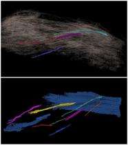
Part of Hughes’ work has focused on creating never-before-seen computer-generated models of this finger muscle. She has been linking cross-sectional images of various parts of the finger, which were captured using the University’s magnetic resonance imaging (MRI) equipment. After transferring the images to a computer, Hughes is able to color in each slide different parts of the muscle, bundles of muscle fibers known as fascicles, and she can track connective tissue through each individual image. Then, she can manipulate those colored slides on a computer, stacking them next to each other as they are positioned in the finger, which results in a 3D model.
Hughes said that seeing muscles in this way is a beneficial supplement to reading about them in her anatomy classes required for the kinesiology major. “I love to see things and know what I’m looking at,” she said.
Hughes helped with one week of data collection early in the summer, during which time participants came into the lab and performed simple finger movement tasks such as wiggling their pointer finger back and forth slowly and contracting finger muscles at different speeds. The majority of her research experience, however, has been analysis. She pores through scientific textbooks and scholarly articles to help make sense of the data. She and Infantolino will spend the entire summer, and perhaps longer, sorting through data collected during that one week.
“This has been a great experience for me. I’ve learned so many things that I can’t count them all,” said Hughes. “I never thought I could or would do research, but now I know that I can.”
Hughes will continue her research into the fall semester and present a synopsis in spring 2011 at the Undergraduate Research Exhibition.


